Ct Neck Radiopaedia . In the emergency setting, a systematic approach to interpreting neck ct findings, including evaluation of findings in the cutaneous and subcutaneous soft tissues, thyroid and. Labeled and unlabelled images of a contrast ct of the neck. The recently inserted thoracic aortic stent proximal margin. No acute intracranial hemorrhage or mass effect. I have uploaded it so that it can be. The dural venous sinuses appear normal. Normal imaging for anatomy reference. Scrollable ct highlighted the anatomy of the. This is a ct scan with contrast of a normal young adult. Patent circle of willis without large vessel. Normal circle of willis and arch anatomy.
from mavink.com
Patent circle of willis without large vessel. Scrollable ct highlighted the anatomy of the. The dural venous sinuses appear normal. Normal circle of willis and arch anatomy. Labeled and unlabelled images of a contrast ct of the neck. No acute intracranial hemorrhage or mass effect. I have uploaded it so that it can be. In the emergency setting, a systematic approach to interpreting neck ct findings, including evaluation of findings in the cutaneous and subcutaneous soft tissues, thyroid and. This is a ct scan with contrast of a normal young adult. Normal imaging for anatomy reference.
Ct Scan Neck Anatomy
Ct Neck Radiopaedia The dural venous sinuses appear normal. This is a ct scan with contrast of a normal young adult. No acute intracranial hemorrhage or mass effect. The recently inserted thoracic aortic stent proximal margin. Labeled and unlabelled images of a contrast ct of the neck. Scrollable ct highlighted the anatomy of the. Normal circle of willis and arch anatomy. In the emergency setting, a systematic approach to interpreting neck ct findings, including evaluation of findings in the cutaneous and subcutaneous soft tissues, thyroid and. I have uploaded it so that it can be. The dural venous sinuses appear normal. Patent circle of willis without large vessel. Normal imaging for anatomy reference.
From www.pinterest.com.mx
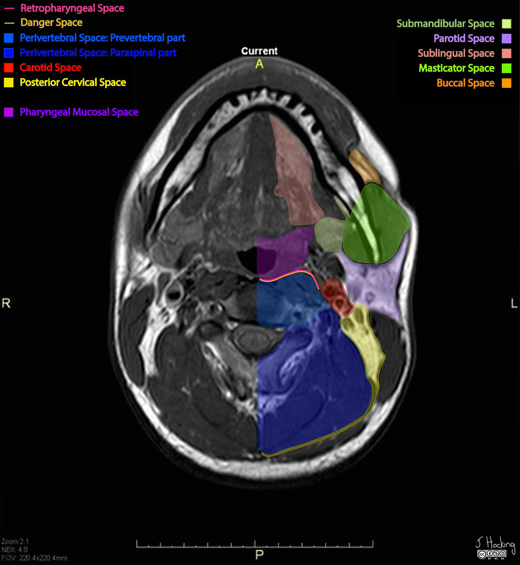
Deep spaces of the head and neck annotated MRI Radiology Case Ct Neck Radiopaedia No acute intracranial hemorrhage or mass effect. I have uploaded it so that it can be. Normal imaging for anatomy reference. Scrollable ct highlighted the anatomy of the. This is a ct scan with contrast of a normal young adult. Normal circle of willis and arch anatomy. In the emergency setting, a systematic approach to interpreting neck ct findings, including. Ct Neck Radiopaedia.
From radiopaedia.org
Normal CT of the neck Image Ct Neck Radiopaedia No acute intracranial hemorrhage or mass effect. In the emergency setting, a systematic approach to interpreting neck ct findings, including evaluation of findings in the cutaneous and subcutaneous soft tissues, thyroid and. The recently inserted thoracic aortic stent proximal margin. I have uploaded it so that it can be. Labeled and unlabelled images of a contrast ct of the neck.. Ct Neck Radiopaedia.
From orlandoappliances4less.com
The Radiology Assistant Cervical Lymph Node Map (2024) Ct Neck Radiopaedia Normal imaging for anatomy reference. This is a ct scan with contrast of a normal young adult. The recently inserted thoracic aortic stent proximal margin. No acute intracranial hemorrhage or mass effect. In the emergency setting, a systematic approach to interpreting neck ct findings, including evaluation of findings in the cutaneous and subcutaneous soft tissues, thyroid and. Labeled and unlabelled. Ct Neck Radiopaedia.
From www.youtube.com
ct scan of neck study part YouTube Ct Neck Radiopaedia Normal circle of willis and arch anatomy. No acute intracranial hemorrhage or mass effect. Labeled and unlabelled images of a contrast ct of the neck. I have uploaded it so that it can be. The recently inserted thoracic aortic stent proximal margin. The dural venous sinuses appear normal. In the emergency setting, a systematic approach to interpreting neck ct findings,. Ct Neck Radiopaedia.
From ottinf.com
The Radiology Assistant Cervical Lymph Node Map (2023) Ct Neck Radiopaedia Normal imaging for anatomy reference. Labeled and unlabelled images of a contrast ct of the neck. Normal circle of willis and arch anatomy. This is a ct scan with contrast of a normal young adult. The dural venous sinuses appear normal. I have uploaded it so that it can be. No acute intracranial hemorrhage or mass effect. Patent circle of. Ct Neck Radiopaedia.
From radiopaedia.org
Image Ct Neck Radiopaedia No acute intracranial hemorrhage or mass effect. This is a ct scan with contrast of a normal young adult. The recently inserted thoracic aortic stent proximal margin. Scrollable ct highlighted the anatomy of the. Labeled and unlabelled images of a contrast ct of the neck. Normal circle of willis and arch anatomy. I have uploaded it so that it can. Ct Neck Radiopaedia.
From www.pinterest.ph
Deep spaces of the head and neck Radiology Reference Article Ct Neck Radiopaedia This is a ct scan with contrast of a normal young adult. The recently inserted thoracic aortic stent proximal margin. Normal circle of willis and arch anatomy. The dural venous sinuses appear normal. Labeled and unlabelled images of a contrast ct of the neck. Normal imaging for anatomy reference. Scrollable ct highlighted the anatomy of the. Patent circle of willis. Ct Neck Radiopaedia.
From www.pinterest.com
Thoracic lymph node stations (annotated CT) Radiology Case Ct Neck Radiopaedia I have uploaded it so that it can be. Patent circle of willis without large vessel. Normal imaging for anatomy reference. Normal circle of willis and arch anatomy. This is a ct scan with contrast of a normal young adult. Labeled and unlabelled images of a contrast ct of the neck. Scrollable ct highlighted the anatomy of the. No acute. Ct Neck Radiopaedia.
From www.ctisus.com
Normal CTA of the Neck Neuro Case Studies CTisus CT Scanning Ct Neck Radiopaedia The recently inserted thoracic aortic stent proximal margin. Normal imaging for anatomy reference. This is a ct scan with contrast of a normal young adult. Normal circle of willis and arch anatomy. Scrollable ct highlighted the anatomy of the. The dural venous sinuses appear normal. Labeled and unlabelled images of a contrast ct of the neck. No acute intracranial hemorrhage. Ct Neck Radiopaedia.
From radiopaedia.org
Gallbladder hydrops Image Ct Neck Radiopaedia The recently inserted thoracic aortic stent proximal margin. No acute intracranial hemorrhage or mass effect. In the emergency setting, a systematic approach to interpreting neck ct findings, including evaluation of findings in the cutaneous and subcutaneous soft tissues, thyroid and. Labeled and unlabelled images of a contrast ct of the neck. Scrollable ct highlighted the anatomy of the. I have. Ct Neck Radiopaedia.
From www.vrogue.co
Ct Neck Axial Anatomy Anatomy Of The Neck Radiology S vrogue.co Ct Neck Radiopaedia The dural venous sinuses appear normal. Normal imaging for anatomy reference. Scrollable ct highlighted the anatomy of the. This is a ct scan with contrast of a normal young adult. Patent circle of willis without large vessel. I have uploaded it so that it can be. The recently inserted thoracic aortic stent proximal margin. Labeled and unlabelled images of a. Ct Neck Radiopaedia.
From nz.pinterest.com
Pin on MSK Ct Neck Radiopaedia This is a ct scan with contrast of a normal young adult. I have uploaded it so that it can be. The recently inserted thoracic aortic stent proximal margin. Normal circle of willis and arch anatomy. Normal imaging for anatomy reference. Scrollable ct highlighted the anatomy of the. The dural venous sinuses appear normal. Labeled and unlabelled images of a. Ct Neck Radiopaedia.
From radiopaedia.org
Image Ct Neck Radiopaedia No acute intracranial hemorrhage or mass effect. The recently inserted thoracic aortic stent proximal margin. I have uploaded it so that it can be. In the emergency setting, a systematic approach to interpreting neck ct findings, including evaluation of findings in the cutaneous and subcutaneous soft tissues, thyroid and. Normal imaging for anatomy reference. Normal circle of willis and arch. Ct Neck Radiopaedia.
From www.pinterest.com
Viewing playlist AAAMARCH Radiology, Mri, Head Ct Neck Radiopaedia Normal circle of willis and arch anatomy. The dural venous sinuses appear normal. I have uploaded it so that it can be. Labeled and unlabelled images of a contrast ct of the neck. No acute intracranial hemorrhage or mass effect. Normal imaging for anatomy reference. This is a ct scan with contrast of a normal young adult. Patent circle of. Ct Neck Radiopaedia.
From www.semanticscholar.org
Figure 5 from Computed tomography imaging of deep neck space infections Ct Neck Radiopaedia I have uploaded it so that it can be. In the emergency setting, a systematic approach to interpreting neck ct findings, including evaluation of findings in the cutaneous and subcutaneous soft tissues, thyroid and. Scrollable ct highlighted the anatomy of the. This is a ct scan with contrast of a normal young adult. Normal circle of willis and arch anatomy.. Ct Neck Radiopaedia.
From www.vrogue.co
Petrous Temporal Bone vrogue.co Ct Neck Radiopaedia Normal imaging for anatomy reference. The dural venous sinuses appear normal. No acute intracranial hemorrhage or mass effect. The recently inserted thoracic aortic stent proximal margin. Patent circle of willis without large vessel. This is a ct scan with contrast of a normal young adult. Labeled and unlabelled images of a contrast ct of the neck. Normal circle of willis. Ct Neck Radiopaedia.
From radiopaedia.org
Lymph node levels of the head and neck (annotated CT) Image Ct Neck Radiopaedia Patent circle of willis without large vessel. In the emergency setting, a systematic approach to interpreting neck ct findings, including evaluation of findings in the cutaneous and subcutaneous soft tissues, thyroid and. The recently inserted thoracic aortic stent proximal margin. Scrollable ct highlighted the anatomy of the. The dural venous sinuses appear normal. Normal imaging for anatomy reference. Normal circle. Ct Neck Radiopaedia.
From radiopaedia.org
Image Ct Neck Radiopaedia Patent circle of willis without large vessel. Normal imaging for anatomy reference. Scrollable ct highlighted the anatomy of the. In the emergency setting, a systematic approach to interpreting neck ct findings, including evaluation of findings in the cutaneous and subcutaneous soft tissues, thyroid and. This is a ct scan with contrast of a normal young adult. The recently inserted thoracic. Ct Neck Radiopaedia.